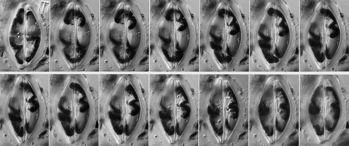
This series of images was taken in 1982. In the first image (top
left), two paired cells of Cymbella
neolanceolata
lie side-by-side in a capsule of mucilage (the
paler, more homogeneous halo around the cells); each contains two
gametes, one above the other, separated by a 'horizontal line'. Then
the gametes fuse with each other and move as a coordinated
pair (clockwise) so that, finally (bottom right image), each
gametangium
contains a single elongate zygote. The black strips inside the cells
are the chloroplasts, which become folded during gametogenesis (which
precedes the stages shown here). This series complements the
information given in a classic early paper on diatom meiosis and
auxosporulation by Lothar Geitler (1927).
Click on the image for a larger version.