Cell-cycle movements of the chloroplast:
summary
Introduction
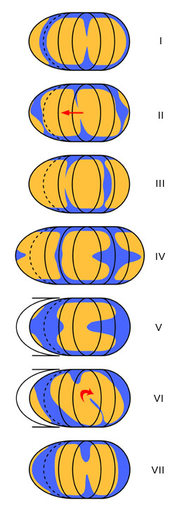
These diagrams summarize the rearrangements of the
chloroplast during the cell cycle in Sellaphora. Each
picture is an equal-area projection of the cell periphery - as if the
cell were stretched out flat laterally. The central ellipse is the
epivalve and this
is flanked by two curved strips representing the two sides of the
girdle. In order to show the hypovalve adequately, the half shown on
the right is duplicated on the left (beyond the
dashed line). The mapping is summarized in a separate diagram.
Description
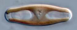
During interphase
(I) the
H- shaped chloroplast lies against the epivalve.
In preparation
for mitosis (II),
the chloroplast moves onto girdle (essentially a translational
movement).
During mitosis
(III), the chloroplast lies with its major constriction
lying along the mid-line of the girdle. This constriction deepens and
cuts the chloroplast in two, just before cytokinesis. The distal side
of the chloroplast, lying on the opposite side of the chloroplast,
develops a wide invagination. At cytokinesis (IV),
the invagination is deep, almost reaching the centre of the epivalve.
After cytokinesis and during
valve formation (V), a second indentation of the
chloroplast begins to develop on the opposite side from the first.
On completion
of the new valve (VI), the chloroplast rotates clockwise
(as seen from the cell exterior) through
90º and the second invagination continues to deepen.
In newly
separated
cells (VII), the chloroplasts are often still slightly
asymmetrical, reflecting the asynchronous development of the two
invaginations.
Interpretation
The chloroplast invaginations that develop during mitosis and
early
interphase represent the first phase of chloroplast division, but
division arrests at a late stage and is not completed until
just
before the next cell division. Similar arrest is seen also in some Caloneis
and Lyrella
species.