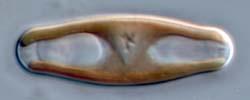
The frustule of Sellaphora
Shown above is the cell wall of a Sellaphora species after treatment with strong oxidizing acids to remove organic material. What is left after oxidation is called the 'frustule', which comprises those parts of the cell wall composed of silica. It consists of two large pieces, called valves, which are connected by a series of narrow, rather flimsy strips of silica called girdle bands. In the photograph one of the two valves is visible; the frustule is resting on the other. Around the valve, you can see the girdle bands, which have become slightly displaced.
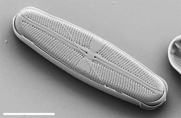
The valve has the same basic structure as most other 'raphid' diatoms: along its midline there is a raphe system, containing two slits that are involved in cell motility, and from here lines of pores extend out more or less perpendicularly. The lines of pores (striae) are separated by narrow ribs of silica. The scale bar represents 10 µm.
The next pages will deal with frustule structure in more detail. Further information is given by Mann (1989) and Round et al. (1990).
As in most other diatoms, the cell wall of Sellaphora species is a composite of amorphous hydrated silica (the product of a partial condensation of orthosilicic acid) and various organic components (proteins, oligo- and poly-saccharides, etc). No analyses of the organic components are available for Sellaphora: they have been studied in detail in only a few diatoms, such as Melosira nummuloides, some Cyclotella species, 'Navicula pelliculosa', or Cylindrotheca fusiformis, although these do in fact represent a wide cross-section of the phylogenetic diversity among diatoms. The article by Hecky et al. (1973) is a good starting point for those interested in the biochemical composition of the organic components of diatom cell walls, but there have been significant advances in the last decade, with the discovery of further classes of organic components involved in silicification and maintaining the integrity of the cell wall (e.g. silaffins and pleuralins: Kröger et al. 2000, Kröger & Wetherbee 2000).