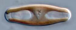
Sellaphora capitata
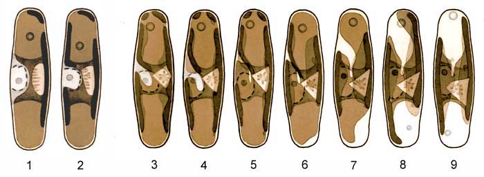
1, 2. Immediately after mitosis and cytokinesis
These are Figs 6 and 7 shown on the 'Premitotic changes' page. Each cell has inherited a single plate of the parent chloroplast; this plate is asymmetrical and becomes even more so as the lateral invagination (at centre-left) deepens and narrows during the formation of the new hypovalve. The pyrenoid is usually rounded during mitosis and cytokinesis (Fig. 1) but soon begins to regain its normal angular shape (Fig. 2). At this stage, the newly formed daughter cell contains one volutin granule (in the top vacuole), which it has inherited from the parent cell. The nucleus is still positioned where it was for mitosis, just beneath the girdle on one side of the cell.
3-9. Post-cytokinetic rearrangements of the protoplast
The chloroplast rotates through 90º. At the same time, the central invagination that had begun to develop before mitosis, on the side of the chloroplast lying beneath the epivalve. During rotation, the apex of the invagination appears to remain physically anchored near the centre of the epivalve (Figs 5-9). The opposite margin of the chloroplast, lying along the girdle close to the plane of division, is at first ± planar but begins to develop an invagination of its own as the chloroplast rotates. However, its development always lags behind the first-formed (epivalvar) invagination (Figs 7-9), so that the chloroplast remains asymmetrical until after rotation is complete. The development of the two invaginations creates two large lobes linked by a narrow isthmus, which become the girdle-appressed plates of the mature H-shaped chloroplast.
The nucleus is displaced inwards as one chloroplast lobe pushes between it and the cell wall (Figs 3-6; compare Fig. 2). A single nucleolus is present throughout interphase. As during interphase, the nucleus is surrounded by a shell of Golgi bodies (dictyosomes), which appear in optical section as short curved bars (creating the 'dashed outline' shown in the drawings).
A second, tiny volutin granule appears during rotation of the chloroplast (Figs 8, 9) and grows until it is ± equal in size to the granule inherited from the parent cell, which lies in the other of the two polar vacuoles.
Finally, the cell regains bilateral and bipolar symmetry, as in the photograph in the header of this page.
Rotation and morphogenesis of the chloroplast take place almost entirely while the daughter cells are still attached to each other follwoing cytokinesis. Separation of the daughter cells occurs while they are in stages corresponding to Fig. 9, or even later, when full symmetry has been achieved.
Cell-cycle changes in chloroplast shape and positions are summarized on a separate page.