 MICROSCOPY
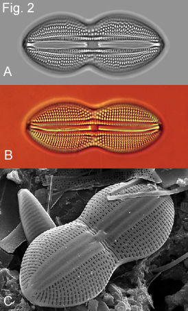
Preparation of diatoms for light microscopy involves cleaning with acid which causes frustules to disintegrate into their component parts (see detailed protocol), these are allowed to settle onto a coverslip which is then permanently mounted onto a slide using a high-refractive-index mountant. Because of their shape, most valves settle in what is called valve view. It is this orientation that is commonly used for identification. Diatoms can be examined using either light or electron microscopy. For the purposes of image analysis or automatic identification brightfield light microcopy (Fig. 2A) is the preferred option as it avoids the shading effects generated by interference contrast (Fig. 2B) and the perspective effects of scanning electron microscopy (Fig. 2C).
![]()