 Image:
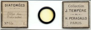
stained slides of
Image:
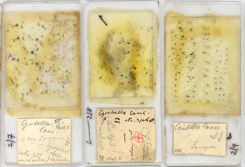
stained slides of Introduction
Professor Dr. Lothar Geitler's research career spanned more than 65 years. His first paper, on blue-green algae (cyanobacteria), was published in 1921, and his last, a nomenclatural paper on the typification of a diatom, in 1987. Altogether, he published 359 papers, monographs and books (a full list was compiled by Dr A.-M.M. Schmid and published in Diatom Research 6: 181–193), dealing with various groups of algae, fungi and lichens, and the cytology of angiosperms and invertebrates. The slides used for his published research appear to have survived largely intact and they are now held in the herbarium of the University of Vienna. In June 2009, I had the opportunity to examine the slide collection and make a preliminary catalogue of the many slides of stained or cleaned diatoms. Geitler's legacy also contains the original illustrations and photographs for most of his published work and a few unpublished notes. However, few significant notebooks are present in the collection and the microscope Geitler used was not found, making it impossible to use the slide locations recorded on some slide labels and notes.
The physical characteristics of the slide collection
The slides are currently contained in over 20 boxes, most of which have a double row of slots and would generally be used to house 200 slides. However, in many cases, slots are occupied by two slides, placed back-to-back. Most slides are of standard size (c. 75 × 25 mm), but there are also some larger slides (c. 75 × 35 mm), kept in separate boxes, which were used for serial sections of Phragmites roots bearing epiphytic diatoms and related material. The slides are arranged in date order and slides of diatoms are interspersed with green algae, higher plants, cyanobacteria and other specimens according to Geitler's varying interests and focus at different stages in his career.
Manuscripts and proofs
The proofs of many of Geitler's papers, together with the original pen and pencil drawings and watercolours for the illustrations, have survived and are kept in folders or original envelopes (many of these are themselves of historical interest!). At present, the folders and envelopes are approximately in chronological order. They include proofs and drawings for Geitler's largest single work, the Cyanophyceae volume of Dr L. Rabenhorst's Kryptogamen-Flora von Deutschland, Österreich und der Schweiz [volume 14, published 1932] and for his pivotal work on the diatom life cycle [L. Geitler (1932). Der Formwechsel der pennaten Diatomeen. Archiv für Protistenkunde 78: 1–226].
Extra drawings and notes
Whether Geitler habitually used notebooks is now unclear and none are present in the collections in the University of Vienna, apart from one small notebook from the 1920s containing drawings and observations of Cymbella lanceolata. Some of the microscope slide boxes contained one or a few loose sheets of notes, with quick drawings and the microscope stage coordinates of interesting specimens, and similar sheets are present also in the folders and envelopes. In some cases too, extra sheets of paper or thin card are present in the folders containing the proofs of Geitler's papers, bearing drawings that were not used in the final publication. Others have holes where favoured drawings have been cut out after being worked up for publication.
Photographs
On the whole, Geitler preferred to record the structure of cells in drawings, where information from different focal planes can be combined into one image. However, he did sometimes use photographs, even in papers published in the 1920s. Photographic prints used for publications are present in the manuscript folders, but there are almost no negatives. In a couple of cases, inspection of the photographs shows that they were enhanced.
Acknowledgment
Preparation of this and related web-pages was made possible by research that received support from the SYNTHESYS Project http://www.synthesys.info/ which is financed by European Community Research Infrastructure Action under the FP6 "Structuring the European Research Area" Programme.
David Mann, August 2009