Formation of the initial cell
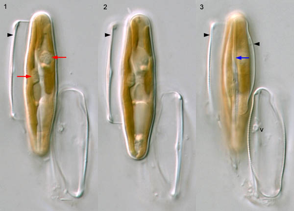
Once the auxospore has finished expanding, the 'initial cell' of the next generation of vegetative cells is formed within the auxospore wall. The example shown is an auxospore in which the epivalve of the initial cell has already been completed: some of its striae and its raphe (Fig. 3, blue arrow) can be seen. At this stage, the auxospore is still closely associated with the gametangia. The male gametangium (lower right in each figure) contains the degenerating remains of the supernumerary cell from meiosis I and of the residual body excluded from the male gamete (one of its volutin granules can be seen in Fig. 3, v). The auxospore's long axis is parallel to the long axis of the female gametangium (arrowheads indicate the displaced thecae of the female gametangium).
At this stage, the auxospore protoplast contains two chloroplasts, one on either side of the cell (Figs 1 and 2), each containing its own pyrenoid (Fig. 1, red arrows): the fingers of cytoplasm that penetrate the pyrenoid appear as small spots or pits. The two gametic nuclei fused earlier, during the later stages of auxospore expansion, and after that the diploid fusion nucleus divided mitotically before formation of the initial epivalve. This mitosis and another that takes place immediately before the formation of the initial hypovalve are not followed by cytokinesis and, at each mitosis, one of the two nuclei produced degenerates. It is a basic rule, very rarely broken, that the formation of each and every valve in diatoms is preceded by a mitosis. In the auxospore shown above, the nucleus is visible just below centre in Fig. 2, lying in a bridge of cytoplasm.