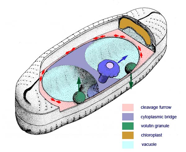
A 3-D reconstruction of the dividing Sellaphora cell
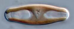
Photographs of dividing cells in girdle and valve views (shown in the previous two pages) led to this attempt at 3-D reconstruction. The action of the contractile ring is indicated by red arrows, the inheritance of the volutin granules by green arrows, and the slight longitudinal displacement of the daughter nuclei (a similar displacement is common in the diatom genera and species we have studied) by a blue arrow, the lower nucleus being displaced in the opposite direction (i.e. the telophase spindle tilts).
This illustration is modified from Mann (1985).