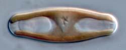
Sellaphora
obesa
cell in valve view, immediately after cytokinesis
Here, a recently divided cell is shown in
valve
view, with the focus exactly in the plane of division. The two daughter
nuclei are not visible, but the two volutin granules are obvious though
just out of focus,
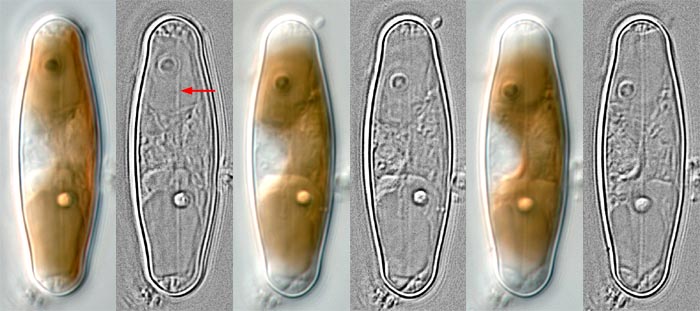
one on either side of the focal plane. Three pairs of photographs are
shown, representing digital exposures at 08.47, 09.11 and
09.30 h.
The cell was first seen at 08.45 h, when a single, straight
longitudinal
element was visible (red arrow); this probably represents the tubular
silicon deposition vesicle. At this stage, it did not lie along the
midline of the cell but slightly to one side.
Later, it became central and by 09.30, regularly spaced lateral
elements could just be detected along the edges of the longtitudinal
element. No raphe slits could be seen at this stage.
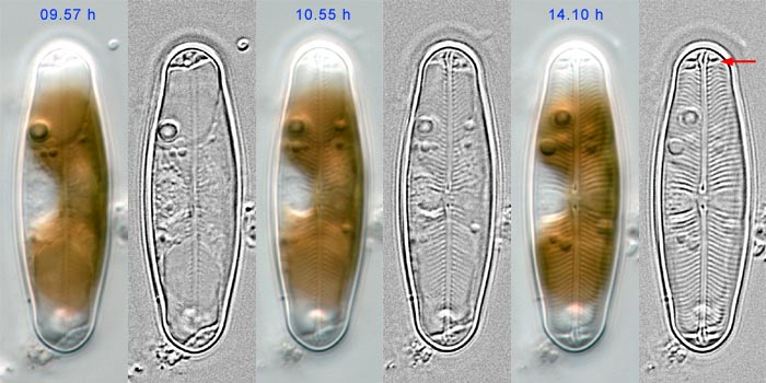
Later stages
in valve formation
The cell was examined at intervals for the next 4.5 hours.
Initially, ribs seemed to be formed along the whole length of
the longitudinal element (the raphe-sternum), including the central
portion. By 10.00 h, however, the distinction between the central ribs
seemed to be breaking down, and by 11.00 h, the plain central area of
the mature valve was obvious. The ribs steadily grew
outwards to the margins to form the transverse ribs of the valve
and thickened, again from the centre outwards. The raphe slits
were visible by 10.00 h and became more and more obvious during the
next 4 hours. The polar bars of the mature valve were visible by 11.00
h, but not fully thickened until later (red arrow). The whole
series of
images from which these three were selected is available for
view; the
image is large and may take some time to load.
The development of Sellaphora
valves has not yet been studied with
electron microscopy. However, it is already clear from light microscopy
that the main
features agree with the classic account of valve formation
in naviculoid diatoms by Chiappino & Volcani (1977).
The original digital photographs (taken using differential
interference contrast optics on a Reichert Polyvar 2 photomicroscope)
were globally enhanced to produce the colour images shown
above,
using the Levels, Brightness/Contrast and Unsharp Mask
tools in Adobe Photoshop. In order to reveal detail of the forming
valves, the original photographs were also reduced to grey-scale images
and processed using a High Pass filter and global
enhancement using
the Auto Levels, Curves and Unsharp Mask tools.