Resolution
go to 'Köhler illumination'Skip to summary
The resolution of an optical system is the smallest distance between two points that can still, just, be seen to be separate. It is the resolution of the microscope that ultimately determines performance. Of course, if the microscope does not also magnify the image, then we will not be able to see the detail that the microscope resolves (because the optics of our eyes and the structure of the retina impose limits on our ability to perceive the detail). However, magnification on its own is worthless. The resolution of the light microscope is limited by diffraction.
The physics of light microscopes was studied in depth in the nineteenth century, especially by Ernst Abbé. He and others (e.g. Lord Rayleigh) established a body of theory and empirical data showing that, for point sources of light in the specimen plane, the resolution (r) of the light microscope is given by the formula
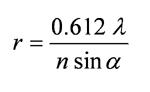
where λ is the wavelength of the light, n is the refractive index of the medium between the point source (specimen) and the objective lens, and α is the 'half-angle of acceptance' of the objective lens, i.e. the angle between the outer limit of the cone of light entering the objective and the optical axis of the lens. The whole quantity {n sin α} is called the numerical aperture (NA) of the objective and is almost always engraved on the lens. The NA of a dry lens cannot be greater than 1, since in that case n = 1 and the sine of no angle is greater than 1. In practice, the very best dry lenses achieve NA 0.95. These have extremely short working distances (the distances between objective and specimen). Most ×40 lenses have NA = 0.65-0.75.
With immersion lenses (immersion oil has a refractive index n = 1.515), much higher NA can be achieved. Student microscopes generally have ×100 lenses with NA = 1.3, while research-quality microscopes often have NA = 1.32 or 1.4.
However, the resolution of real objects in transmitted light is determined not only by the characteristics of the objective, but also by those of the condenser, which after all is providing the cone of light accepted (after modification by the specimen) by the objective. A second formula is necessary:
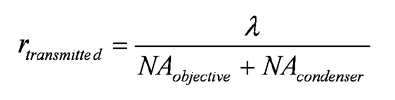
Most people, most of the time, use high-magnification oil immersion lenses with oil between the specimen and the objective, but with air between the specimen and the condenser. In this case, even if the numerical aperture of the condenser is nominally 1.3 (the actual value is usually engraved somewhere on the condenser), its NA will in fact be less than 1 and the NA of the system as a whole will be reduced: a NA 1.3 objective will function more like a NA 1.15 objective.
The higher the numerical aperture, the lower the depth of focus of the specimen. The depth of focus becomes very small with the highest NA lenses. Young et al. [Young, I.T., Zagers, R., van Vliet, L.J., Mullikin, J., Boddeke, F.R. & Netten, H. (1993). Depth-of-focus in microscopy. In: Proc. 8th Scand. Conf. Image Analysis, pp. 493-498. Norweg. Soc. Image Processing and Pattern Recognition, Tromsø, Norway] give:
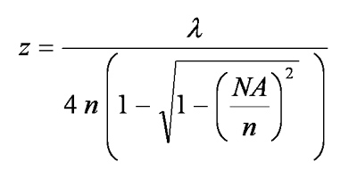
Hence, for a NA = 1.32 objective, used optimally with green light of wavelength 550 nm, the depth of focus is just 0.178 µm.
To summarize:
- the resolution of a light microscope is limited by diffraction
- the higher the numerical aperture of the system, the greater the resolution
- the highest numerical apertures can only be achieved in immersion systems, and for the very best results, the specimen/slide must be oiled not only to the objective but also to the condenser; this, however, can become very messy and it is only too easy to get oil into places in the condenser where it shouldn't be
- achievement of better resolution can be helped by using short wavelength (blue) monochromatic light; but this is hard on the eyes
- with good immersion lenses and properly set-up illumination using green light (550 nm wavelength), a resolution of close to 0.2 µm (200 nm) should theoretically be achievable with a fully immersed system; even if the condenser is not oiled, the resolution should be better than 0.25 µm. In practice, few microscopists achieve better than c. 0.23-0.25 µm, even with the best optics and fully immersed lenses.
- diatomists should note that the theoretical and practical limits of resolution correspond to stria densities of 40-50 in 10 µm. Such densities are sometimes not observed not because of lack of resolution but because the striae are too faint to be focused reliably
- higher resolution in normal, transmitted light microscopy has an inevitable corollary: low depth of focus