Does it matter if the light microscope is not set up correctly?
- What if the condenser diaphragm is closed down too much or not enough?
- Does it matter if the condenser is positioned too low or high?
- Should the condenser be oiled to the slide?
- What about the light?
Condenser position (focus)
back upUnless the condenser is in exactly the right position, Köhler illumination will not be achieved. There is a risk that the cone of light provided by the condenser will not develop (fill) the full aperture of the objective lens, thus compromising resolution, and the illumination will become uneven across the field of view. As with the condenser diaphragm, many people use the wrong settings, lowering the condenser too much, because this seems to give a clearer picture, because the contrast of the specimen is enhanced. Without contrast, nothing can be seen and so it is indeed useful sometimes to use suboptimal settings of the condenser, in order to make observation more compfrtable, especially during long sessions. However, for photomicroscopy, the correct settings should be restored.
In all of the images presented on this page, approximately the same degree of enhancement was applied - the same as was applied during examination of the effects of ondenser aperture alone. In each case, the Levels tool in Photoshop was used to expand the dynamic range of the image to the full range from black to white, followed by further slight enhancement of contrast.
- condenser position and aperture optimal
- condenser low
- condenser very low
- condenser very low, but partial compensation by wide aperture
- condenser extremely low
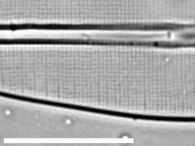
1. Condenser position correct and condenser diaphragm closure optimal, according to the requirements for Köhler illumination.
back up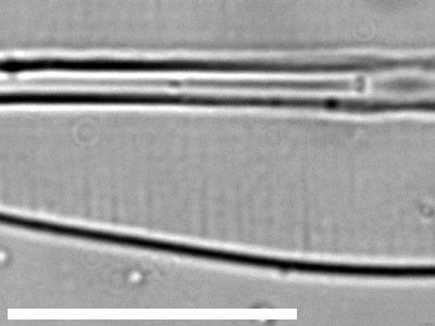
2. Condenser diaphragm left unaltered from 1, but condenser lowered a little. Already, the striae cannot be seen. However, larger structures, such as the ribs bordering the raphe and the valve margin, are still relatively clear. It would be easy to assume that this is all that the microscope could resolve, if image 1 were not available for comparison. Note that many sets of small concentric rings have become visible. These are caused by tiny spots of dust or imperfections on or in the lenses and filters within the microscope; with correct Köhler illumination, most of these would be out of focus.
back up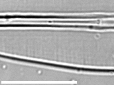
3. Condenser diaphragm left unaltered from 1 and 2, but condenser lowered further. Now even the larger valve structures are becoming fuzzy, but the dust speck artifacts present in 2 have disappeared again, because the condenser is no longer focusing them into the image.
back up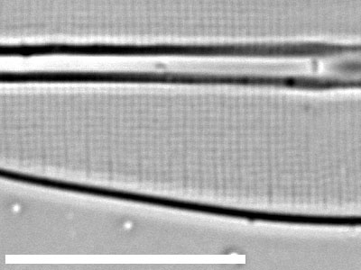
4. Condenser position as in 3 and therefore seriously suboptimal, but with the condenser diaphragm opened fully.
This particular microscope condenser has a sufficiently high aperture that it is possible to compensate to some extent for condenser position by opening the condenser diaphragm. The striae are now visible again, though they are not as clear as with correct Köhler illumination (compare the zone at the top, above the raphe, in 1 and 4)
back up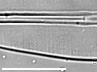
5. Condenser diaphragm fully open, condenser lowered further. Although the raphe ribs and valve margin are still well defined, the striae are invisible. New sets of small concentric rings are visible (especially at bottom right), again caused by dust or imperfections in or on lenses and other glass surfaces within the microscope (note that none correspond to the ring artifacts in 2).
back up