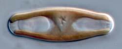
Sellaphora cell in valve view, during cytokinesis
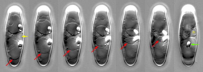
In these photographs, a dividing cell has been photographed in valve view, with the focus exactly in the plane of division. This reveals the shape of the cleavage furrow (red arrows) as it cuts through the cell. In the first frame, the nucleus is still undivided (yellow arrow) in the central cytoplasmic bridge and the furrow has scarcely begun to cut through the polar vacuoles. In the penultimate frame, it reaches the central cytoplasmic bridge containing the mitotic nucleus (which by then will be in telophase). In the last frame, cytokinesis is complete and one of the daughter nuclei is visible (n); for this photograph alone, the plane of focus has been altered slightly, to lie just within the upper daughter cell.
At first, the cleavage furrow does not affect the central part of the cell and the organelles and cytoplasm here are not partitioned. However, it is known from other diatoms that cleavage is achieved through a ± homogeneous 'contractile ring' of microfilaments (F-actin), which is continuous around the perimeter of the cell. The unconstrained shape that would be taken by such a contractile ring cutting through a turgid cell would be a perfect circle, and in centric diatoms the profile of the cleavage furrow is indeed close to being a circle. In elongate diatoms, however, the contractile ring is constrained by the cell wall. Until the ring has a diameter less than the width of the cell, its shape is constrained by the cell wall. The shape is therefore circular only where it is cutting through the protoplast and vacuoles. In the frames shown above, the contractile ring (= the edge of the cleavage furrow) can be seen to be the arc of a circle where the ring is unconstrained by the wall (red arrows). In this Sellaphora, because of the lanceolate shape of the valve, the diameter of the arc is at first small (because the poles are narrow), then increases (as the furrow begins to cut through the wider part of cell), and finally decreases again (as it becomes free from the cell periphery and completes cytokinesis).
Note that the two strongly refractile volutin granules (which are circular but appear elliptical in the frames because of their rapid oscillation and the relatively long exposures used) move inwards as the cleavage furrow develops. In fact, the rate of their inward movement correlates exactly with the contraction of the contractile ring. Finally, the volutin granules are partitioned between the two daughter cells, one into each; this is released into one of the polar vacuoles (last frame).
The central cytoplasmic bridge narrows as cleavage proceeds, but the two vacuoles appear to remain separate.